Discover Cutting-Edge Advanced Microscopy Techniques
- contactelmaslabs
- Sep 6, 2025
- 4 min read

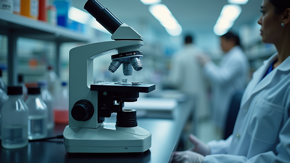
Microscopy has always been a cornerstone of scientific discovery. Today, the field is evolving rapidly with new technologies that allow us to see the tiniest details of life and materials like never before. These innovations are transforming research in biotech companies, academic labs, and startups. If you want to stay ahead, understanding these cutting-edge microscopy methods is essential.
Let me take you through some of the most exciting advances. I’ll explain how they work, why they matter, and how you can apply them to your research. By the end, you’ll feel confident exploring these tools and integrating them into your projects.
Exploring Cutting-Edge Microscopy Methods
Microscopy is no longer just about magnifying objects. Modern techniques combine optics, physics, and computer science to reveal structures at the nanoscale. Here are some of the most impactful methods:
Super-Resolution Microscopy: Breaks the diffraction limit of light to see details smaller than 200 nanometres.
Electron Microscopy: Uses electron beams instead of light for ultra-high resolution images.
Atomic Force Microscopy (AFM): Maps surfaces at the atomic level by “feeling” the sample with a tiny probe.
Light Sheet Microscopy: Illuminates samples with a thin sheet of light for fast, 3D imaging with minimal damage.
Each method has unique strengths. For example, super-resolution microscopy is perfect for studying proteins inside cells, while electron microscopy excels at revealing the fine structure of materials.

These methods are not just fancy gadgets. They provide actionable insights that can accelerate your research. For instance, you can track how a drug interacts with cells in real time or map the surface of a new biomaterial with atomic precision.
How These Cutting-Edge Microscopy Methods Benefit Your Research
Adopting these advanced tools can transform your workflow and results. Here’s how:
Increased Resolution and Detail
You can see structures and interactions that were previously invisible. This leads to more accurate data and better understanding.
Faster Data Acquisition
Techniques like light sheet microscopy allow you to capture 3D images quickly, saving time and reducing sample damage.
Quantitative Analysis
Many modern microscopes come with software that helps you measure and analyze images precisely. This reduces human error and improves reproducibility.
Versatility
These methods can be applied to a wide range of samples - from living cells to complex materials.
Integration with AI
Advanced image analysis powered by AI can automate pattern recognition and data interpretation, making your research more efficient.
By integrating these methods, you can push the boundaries of what’s possible in your experiments. Whether you’re developing new therapies or creating innovative materials, these tools provide a competitive edge.

What are the advanced fluorescence microscopy techniques?
Fluorescence microscopy has been a game-changer in biological research. It uses fluorescent dyes or proteins to label specific parts of a sample, making them glow under certain light. Advanced fluorescence microscopy techniques take this further by improving resolution, speed, and depth.
Some key techniques include:
STED (Stimulated Emission Depletion) Microscopy
This method sharpens images by selectively turning off fluorescence around the focal point, achieving nanoscale resolution.
PALM (Photoactivated Localization Microscopy) and STORM (Stochastic Optical Reconstruction Microscopy)
These rely on switching fluorescent molecules on and off to build super-resolved images from many frames.
Light Sheet Fluorescence Microscopy (LSFM)
Illuminates samples with a thin sheet of light, reducing photodamage and allowing fast 3D imaging of living specimens.
Multiphoton Microscopy
Uses longer wavelengths to penetrate deeper into tissues, ideal for imaging thick samples.
These techniques allow you to observe dynamic processes inside cells with incredible clarity. For example, you can watch how proteins move during cell division or track the development of neurons in real time.

If you want to master these methods, consider workshops and training sessions that focus on image analysis and microscopy. They can help you get the most out of your equipment and data.
Practical Tips for Implementing Advanced Microscopy in Your Lab
Getting started with these cutting-edge methods might seem daunting, but it’s easier than you think. Here are some practical steps:
Assess Your Needs
Identify the questions you want to answer and the samples you work with. This will guide your choice of microscopy technique.
Invest in Training
Attend workshops or partner with experts who can teach you how to operate the instruments and analyze data effectively.
Optimize Sample Preparation
Proper sample handling is crucial for good imaging. Follow protocols carefully to preserve structures and fluorescence.
Leverage Image Analysis Software
Use advanced software tools to quantify and interpret your images. Many platforms now include AI features to speed up analysis.
Collaborate with Specialists
If possible, work with microscopy core facilities or service providers who have experience with these techniques.
By following these tips, you can integrate advanced microscopy into your research smoothly and confidently.
Unlocking the Full Potential of Your Research with Expert Support
To truly benefit from these innovations, expert guidance is invaluable. Partnering with specialists who understand both the technology and your research goals can make a big difference.
At Elmas Labs, we focus on helping you achieve highly precise and reproducible results. Our expertise in image analysis and integration of AI techniques ensures you get the most from your microscopy data. Whether you need training, consultation, or custom solutions, we are here to support your journey.
If you want to explore advanced microscopy techniques in depth, consider joining our image analysis workshops. They are designed to empower you with practical skills and knowledge.

Embracing these cutting-edge microscopy methods will open new doors for your research. With the right tools and support, you can uncover insights that drive innovation and discovery. Let’s take this exciting step forward together!


Comments